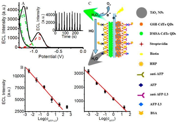
Figure 8.
(A) ECL curves of simultaneous scan of DMSA-CdTe QDs and TiO2-GSH-CdTe QD composites; individual scan of (a) DMSA-CdTe QDs, (b) TiO2-GSH-CdTe QD composites, and (c) modified ITO electrodes. Inset: Continuous cyclic simultaneous scans of DMSA-CdTe QDs and TiO2-GSH-CdTe QD composites modified ITO electrodes. (B) Linear calibration plots for detection of AFP-L3 (left) and AFP (right). (C) Sandwich immuno-structure of HRP-labeled antibody–antigen biotinylated antibody on the immunosensor and detection procedures of analytes (green arrow: luminescence; gray arrow: quenching). Reproduced from [66] with permission from the American Chemical Society.

