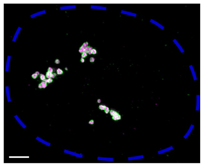
Fig. 1. Super-resolution image of paraspeckle nuclear bodies constructed by NEAT1_2 lncRNA.
Fluorescence in-situ hybridization was performed on HeLa cells to visualize NEAT1_2 5′ and 3′ regions (shown in green) and NEAT1_2 middle region (shown in magenta). A maximum intensity projection image of one nucleus, delineated by a blue dotted line, is shown. Scale bar, 2 μm. Each paraspeckle is estimated to contain about 50 NEAT1_2 molecules in HeLa cells (Chujo et al., 2017). Three clusters of paraspeckles might correspond to the approximate locations of three NEAT1 loci on three copies of chromosome 11 in HeLa cells. 5′ and 3′ regions of NEAT1_2 are located at the “shell” of paraspeckles and the middle region is located at the “core”, as previously reported (Souquere et al., 2010; West et al., 2016).

