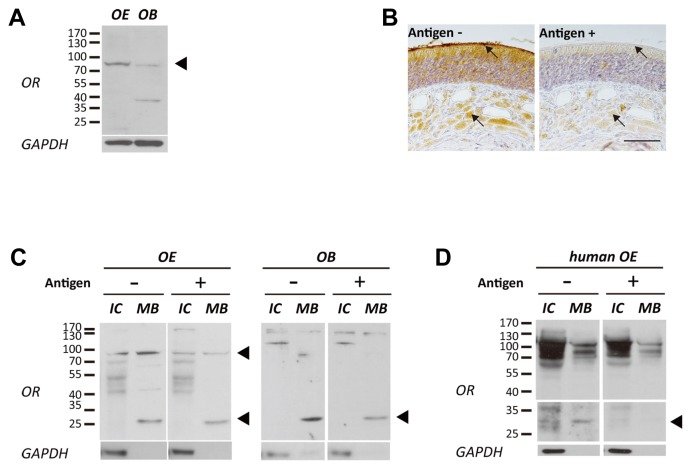
Fig. 3. Expression pattern of PMLNPFIY motif conserved OR in vivo tissues.
(A) The ORs were specifically detected at the size of over 70 kDa in whole lysate of rat OE and OB (Black arrow head indicated). (B) Histological expression pattern of ORs that having target sequences. ORs were observed at the olfactory cilia and nerve bundles of OE tissue (Black arrow points cilia and nerve bundle, Left panel). The specific signals were disappeared after peptide blocking (Antigen conc.;100 ng/ml; Right panel; Scale bar represents 50 μm). (C) Membrane bound (MB) proteins of rat OE and OB were fractionated from intracellular (IC) proteins. Expression of ORs at each sample was confirmed with CAS-TM7 OR antibody which is pre-blocked with or without antigen (Antigen conc.; 1 ng/ml). The intensity of OR specific bands were decreased after pre-incubated with antigen peptide (VTPMLNPFIYSLRNRDC) (Black arrow heads indicate decreased OR bands at OE and OB). (D) Fractionated membrane bound (MB) proteins of human OE also showed expression of ORs at 25~35 kDa and over 70 kDa, and these bands became smear after incubation with peptide pre-blocked CAS-TM7 OR antibody (Antigen conc.; 1 ng/ml).

