
Figure 3.
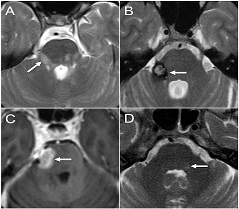
Brainstem lesions causing TN. (a) Axial T2-weighted image through the brainstem demonstrates multiple hyperintense lesions in the posterior fossa in MS including a lesion extending along the expected course of the fascicular fibers and towards the root entry zone of the right CN V (arrow). (b) Axial T2-weighted image through the brainstem shows a characteristic cavernous malformation with central hyperintesities and surrounding hypointense hemosiderin rim (arrow). The lesion is centered in the right side of the pons extending towards the root entry zone of the right CN V. The patient presented with right-sided TN. (c) Axial postcontrast T1-weighted image shows abnormal enhancing lesion extending from the right side of the pons (arrow) into the cisternal segment of the right CN V. Biopsy revealed primary CNS lymphoma. (d) Axial T2-weighted image through the pons shows asymmetric subtle hyperintense signal along the cranial end of the trigeminal nucleus of CN V (arrow). The patient presented with left facial shingles and pain, and was diagnosed with trigeminal zoster.