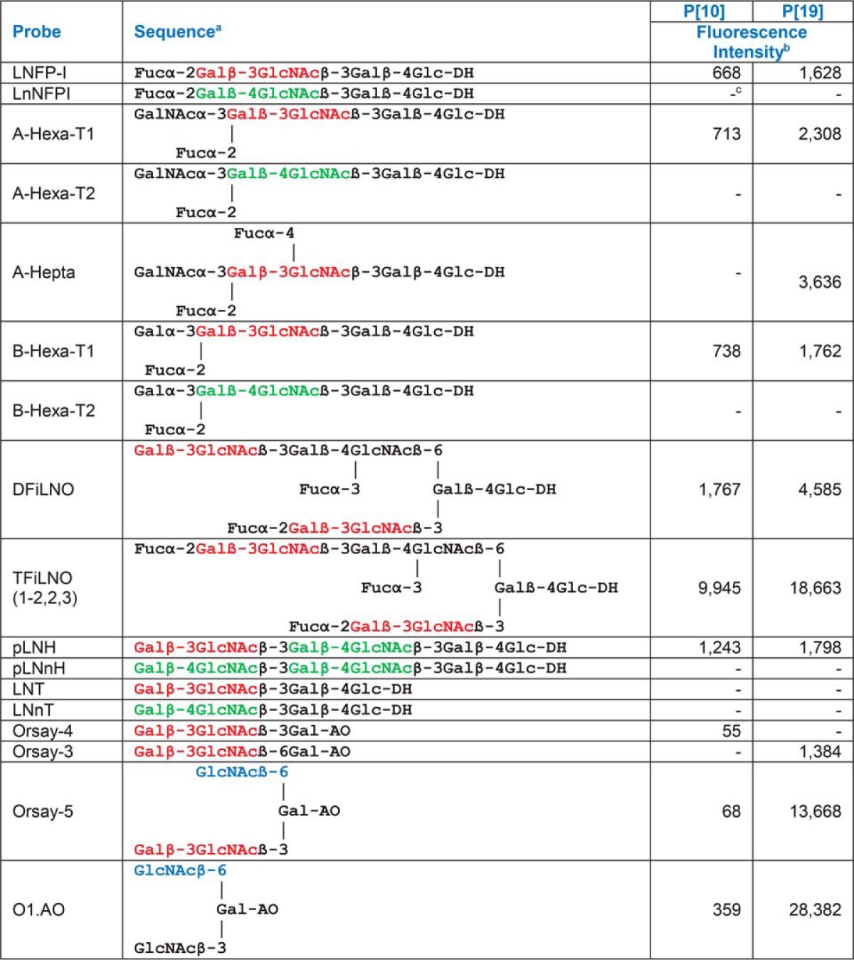
Table I. Fluorescence intensities in microarray analyses of the VP8* proteins of P[10] and P[19] (analyzed at 50 μg/ml) to probes selected for comparison in the F77/Ii array. Sequences with type 1 (Galβ1–3GlcNAc) and type 2 (Galβ1–4GlcNAc) terminating backbones, and terminal GlcNAc are shown in red, green, and blue font, respectively.

a DH-NGLs were prepared from reducing oligosaccharides by reductive amination with the amino lipid, 1,2-dihexadecyl-sn-glycero-3-phosphoethanolamine (DHPE); AO-NGLs were prepared from reducing oligosaccharides by oxime ligation with an aminooxy (AO) functionalized DHPE (40).
b Fluorescence intensities with probes printed at 5 fmol per spot.
c − Indicates fluorescence intensity less than 1.
