Fig. 5.
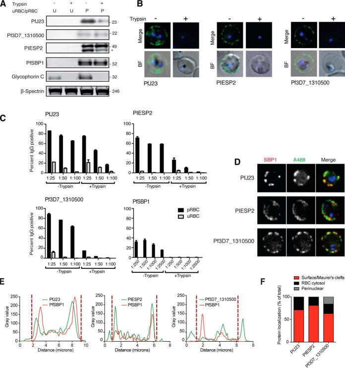
Validation of candidates. A, Unparasitized RBC and intact and live trophozoite stage pRBC (± pre-treatment with trypsin/chymotrypsin) analyzed by Western blot, and membranes were probed with polyclonal antibodies targeting candidate. A trypsin sensitive band at the expected size was detected for pRBCs probed with PfJ23 and PIESP2, whereas detection of Pf3D7_1310500 did not appear to be trypsin dependent. PfSBP1, Glycophorin C, and β-spectrin were included as controls. Each lane represents protein extract from 2.5 × 10⁶ pRBC. Full Western blots are provided in supplemental Fig. S3. B, Surface reactivity of candidate antibodies to purified pRBCs (± pre-treatment with trypsin/chymotrypsin) was detected by live microscopy. Cells were stained with the nuclear dye Vybrant violet and the surface labeled using AlexaFluor488 goat anti-rat secondary antibody. Antibody signal is only detectable on the pRBC surface without prior trypsin treatment of the cells. C, Surface reactivity of candidate antibodies to purified pRBCs (± pre-treatment with trypsin/chymotrypsin) was detected by flow cytometry. Cells were gated for live cells and single cells, and uRBCs and pRBCs were subsequently separately gated based on Vybrant Violet fluorescence and surface reactivity measured using AlexaFluor488 goat anti-rat secondary antibody. Surface reactivity was measured as percent of cells positive for AlexaFluor488. Surface reactivity of all antibodies tested decreased with trypsin treatment, but this change was most significant for PIESP2, Pf3D7_1310500 and the lowest dilution (1:100) of PfJ23. D, Immunofluorescence analysis of the localization of PfJ23, PIESP2 and Pf3D7_1310500 (detected with specific sera) in fixed and permeabilized trophozoite pRBCs shows partial colocalization in the cytosol of pRBC with the Maurer's cleft resident protein PfSBP1, as well as pRBC surface localization. E, Fluorescence plot profile of PfJ23, PIESP2 and Pf3D7_1310500 localization confirms colocalization with PfSBP1 in addition to surface localization. Dashed lines mark the boundary between Maurer's cleft and peripheral/surface labeling based on SBP1 distribution. F, Immunofluorescence-based analysis of protein localization across multiple cells classified by fluorescence intensity percentage, shows that up to 70% of PfJ23, 90% of PIESP2, and 69% of Pf3D7_1310500, respectively, localizes to the pRBC surface or Maurer's clefts, whereas the remaining localization is cytosolic (and perinuclear in the case of Pf3D7_1310500).

