Figure 3.
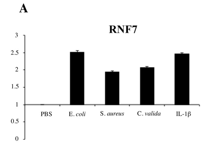
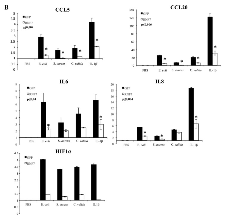
RNF7 expression represses NF-κB signaling upon pathogen-associated molecular pattern (PAMP) recognition. (A) HaCaT cells were left in phosphate-buffered saline (PBS) or exposed to the indicated heat-killed microorganisms or IL-1β (10 ng/mL) for 6 h, and the expression level of RNF7 was monitored by real-time PCR. Graph show the fold changes respect to the cells left in PBS. Data shown is representative of three independent experiments done in triplicate. (B) HaCaT cells were infected with a lentiviral vector expressing RNF7 or control GFP. Forty-eight hours later, cells were left in PBS or exposed to the indicated heat-killed microorganisms for 6 h, and the expression levels of selected NF-κB target genes were monitored by real-time PCR. Data shown represents the fold changes respect to the GFP-transfected cells left in PBS. Data were analyzed by Student’s t-test, and a p-value ≤ 0.05, indicated with an * was considered significant. Data shown is representative of at least three independent experiments done in triplicate.


