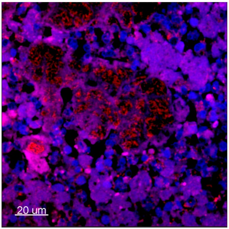
Figure 1.
Confocal laser-scanning microscopy micrography of ex vivo lung tissue from a P. aeruginosa-infected CF patient. Tissue was stained with peptide nucleic acid fluorescence in situ hybridization (PNA-FISH) probes specific for P. aeruginosa with a red Texas-Red flourophor and counterstained with blue (4′,6-diamidino-2-phenylindole) DAPI for eukaryotic nucleus. 630×. [19].

