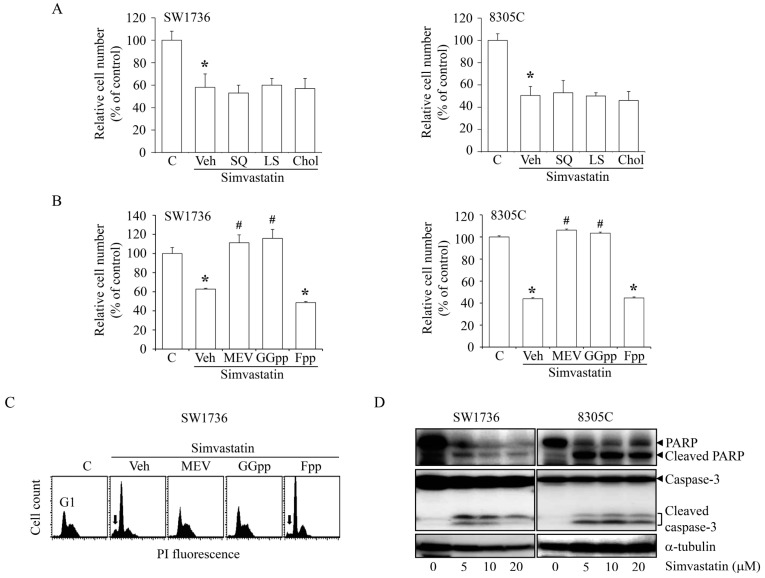
Figure 2.
Effects of MEV-derived metabolites on the simvastatin-induced ATC cell proliferation inhibition. (A) Simvastatin (10 μM)-inhibited proliferation of SW1736 and 8305C cells was not affected by the add-in SQ (10 μM) or LS (10 μM) or Chol (10 μM). Cells were pre-incubated with SQ, LS or Chol for 30 min followed by simvastatin for additional 48 h. The simvastatin (10 μM) induced cell proliferation inhibition (B) and accumulation of cells at the G1-phase (C) of SW1736 and 8305C cells were abolished by MEV (50 μM) and GGpp (20 μM), but not Fpp (20 μM). Cells were pre-incubated with MEV, GGpp or Fpp for 30 min followed by simvastatin for additional 48 h. The relative cell number was estimated using MTT assays. Values represent the means ± SEM (n = 3). The DNA content was measured by PI staining. Arrows indicated sub-G1 cell population. (D) Simvastatin triggered apoptosis in SW1736 and 8305C cells. Cells were incubated with 0–20 μM simvastatin for 48 h, and then the protein expression levels of full-length and active form of caspase 3 and PARP were examined by immunoblotting analysis. * p < 0.05, different from corresponding control. # p < 0.05, different from simvastatin-incubated group. C, control; Veh: simvastatin-incubated group.

