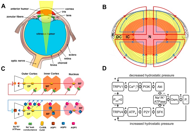
Figure 1.
Structure and function of the ocular lens. (A) The cornea and the lens work together to focus light onto the retina. In the young primate lens the contraction of the ciliary muscle can alter the tension applied to the lens via the zonular fibers and change the shape and therefore optical power of the lens. This process of accommodation is lost as the lens becomes progressively stiffer through middle age resulting in presbyopia. With advanced age, the lens gradually loses it transparency which can ultimately manifest as cataract. Adapted from Wikimedia Commons contributors. Three Internal Chambers of the Eye.png. Available online https://commons.wikimedia.org/w/index.php?title=File:Three_Internal_chambers_of_the_Eye.png&oldid=223135955 (accessed on 10 November 2017); (B) the lens is composed of a single layer of epithelium (E) located at the anterior pole and elongated fiber cells that fill the bulk of the lens. At a narrow region around the equator the epithelial cells undergo differentiation and transform into fiber cells that ultimately become organized into symmetrical growth sheets that stay viable throughout one’s life. The older cells are located in the center or nucleus (N) of the lens while the newly differentiated fiber cells are found in the periphery or outer cortex (OC) of the lens. A net flux of Na+ and fluid enters the lens at both poles and exits at the equator to generate an internal microcirculatory system that delivers nutrients to and removes waste products from the avascular lens faster than would be achieved by passive diffusion alone; (C) spatial differences in ion transport properties generate the microcirculation. Top panel: Na+ flows into the lens along the extracellular spaces between cells and crosses fiber cell membranes by diffusing down its electrochemical gradient, then flows back to the lens surface via an intracellular pathway mediated by gap junctions that direct Na+ to the equator where the Na+/K+ ATPase transports it out of the lens to complete the circulation; Bottom panel: Water follows the circulating Na+ current entering the lens at each pole via the extracellular space before entering fiber cells through AQP0/5 water channels before flowing back to the surface via gap junctions and leaving the lens through AQP1 channels located in the epithelium; (D) the movement of water through gap junctions generates a hydrostatic pressure gradient (Pi, mmHg) that helps to drive the water from the core to the surface. This hydrostatic pressure gradient is dynamically regulated by a dual feedback system that uses the mechano-senstiveTRPV1 and TRPV4 to sense increases and decreases in pressure, respectively and to activate signaling pathways that reciprocally alter Na+, K+-ATPase activity to maintain a constant pressure.

