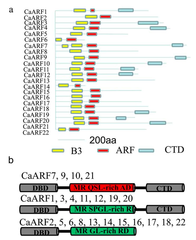
Figure 3.
Analysis of ARF protein structures and domains. (a) Depiction of the domain structure of each CaARF protein sequence. The B3 DNA bindingdomain (BDB), auxin response factor domain (ARF) and Carboxy-Terminal Domains (CTD) are colored in yellow, red and light blue, respectively. (b) Three kinds of CaARF protein structures. DBD, DNA-binding domain; CTD, C-terminal dimerization domain; MR, middle region; RD, repression domain, showed in green color ; AD, activation domain, showed in red color; Q, glutamine; S, serine; L, leucine; P, proline; G, glycine.

