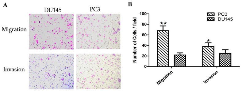
Figure 1.
Comparison of migration and invasion capacity of prostate cancer cells. (A) Representative images of the invasion and migration of DU145 and PC3 cells taken by an inverted microscope (20× objective); (B) Quantitative analysis of cell migration (2 h) and invasion (4 h) in DU145 and PC3 cells. * p < 0.05 ** p < 0.01. Per condition, three independent experiments were performed.

