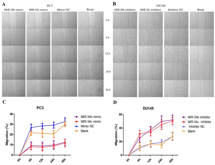
Figure 6.
Random migration by a wound healing/scratch assay. (A,B) Shown are representative images of the scratch assay in DU145 and PC3 cells at 0, 6, 12, 24 and 36 h after the scratch was created; (C,D) The rate of migration calculated by the width of the wound in DU145 and PC3 cells after ectopic expression of miR-34b/c by Image-Pro Plus software (Media Cybernetics, MD, USA).

