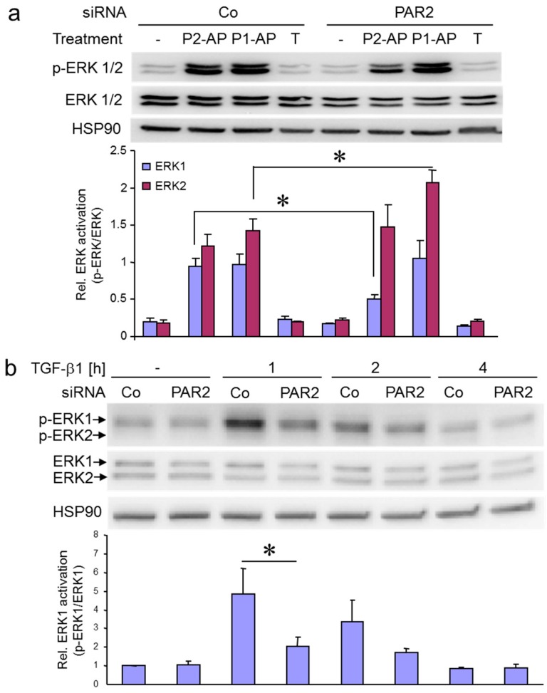
Figure 4.
Both PAR2–AP- and TGF-β1-induced ERK activation are dependent on PAR2 protein expression. (a) Panc1 cells were transiently transfected with 50 nM of either control (Co) siRNA or siRNA specific to PAR2 (PAR2). Following stimulation with PAR2–AP (P2-AP), PAR1–AP (P1-AP) for 5 min or TGF-β (T) for 1 h, cells were subjected to immunoblotting for p-ERK1/2 and ERK1/2, and for HSP90 as a loading control. The graph below the blot shows densitometric data (mean ± SD) of underexposed bands derived from three parallel wells. One representative experiment is shown out of three performed in total. Asterisks indicate significance p < 0.05; (b) HaCaT cells were transfected with 50 nM of either Co siRNA or PAR2 siRNA, stimulated for the indicated times with TGF-β1, and processed for immunoblotting of p-ERK1/2 and ERK1/2. The graphs below the blots show results from densitometry-based quantification of three experiments, mean ± SD. The asterisk indicates significance p < 0.05.

