
Figure 1.
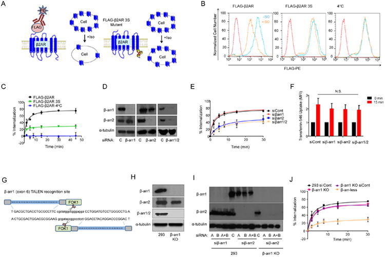
Internalization of β2AR in 293, β-arr1 KO and β-arr-less cells. A) Schematic of FLAG-β2AR and FLAG-β2AR 3S internalization assays. B) Surface FLAG-β2AR or FLAG-β2AR 3S abundance in 293 cells before or after 15 min Iso (10 μM) stimulation compared with isotype-stained reference control, and control stimulation at 4°C. C) Internalization of FLAG-β2AR or FLAG-β2AR 3S after Iso stimulation; mean of three independent experiments +/- standard deviation (SD). D) Western blot of β-arr1 and β-arr2, and a-tubulin (loading control), in 293 cells transfected with β-arrestin or control siRNAs. Representative of four independent experiments. E) Internalization of FLAG-β2AR after Iso stimulation in 293 cells transfected with β-arrestin or control siRNAs; mean of three independent experiments +/- SD. F) Flow cytometry quantification of MFI of 15 min transferrin-546 uptake in 293 FLAG-β2AR cells transfected with control siRNA (siCont) or indicated siRNAs; mean of three independent experiments +/- SD. G) Schematic of the β-arr1 TALEN construct design targeting exon 6. (H and I) Western blot for β-arrestins in (H) 293 and β-arr1 KO cells and in (I) 293 and β-arr1 KO cells transfected with two different siRNAs (A, B) targeting β-arr1 or β-arr2, or siRNA pools (A+B). J) Internalization of FLAG-β2AR after Iso stimulation in indicated cells, mean of three independent experiments +/- SD.