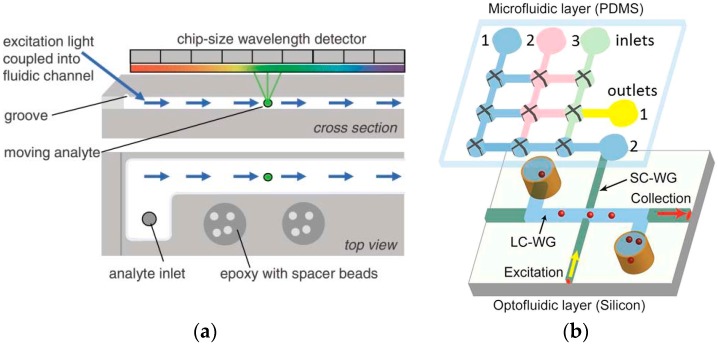
Figure 4.
(a) The cross section and top view of the fluidic channel. The excitation light is coupled into the fluidic channel and the imager records the fluorescence emission of the analytes while they move along the channel. Reproduced from [63] with permission from The Royal Society of Chemistry. (b) The prototype of the ARROW chip for sample preparation and single nucleic acid measurement. Reproduced from [64] with permission from The Creative Commons Attribution 4.0 International License.

