Fig. 2.

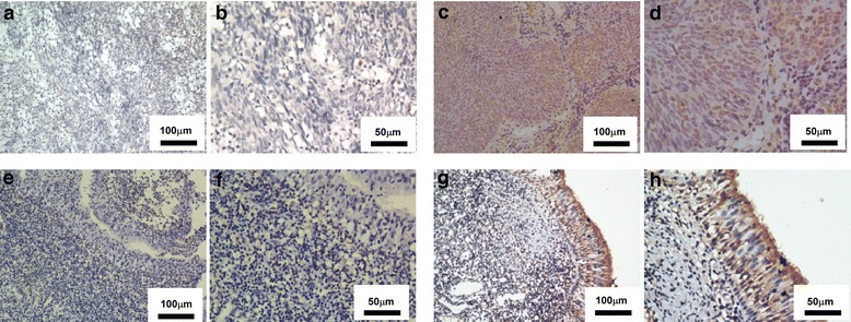
Expression of CCL27 in nasopharyngeal epithelial tissue from a NPC patient and a VCA-IgA–positive healthy donor. a–h Representative images showing immunohistochemical staining of CCL27 in NPC tumor tissue (low CCL27 expression: a-b, high CCL27 expression:c-d) and VCA-IgA–positive healthy tissue (low CCL27 expression: e-f, high CCL27 expression: g-h). Scale bars: a and c = 100 μm; b and d = 50 μm
