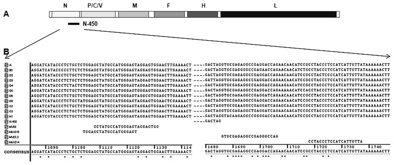
Fig. 1.
Genetic variability of primer binding sites among 11 genotypes. (A) Schematic representation of measles genome and location of N-450. N, P/C/V, M, F, H, L are measles genes. (B) Nucleotide sequence of primer binding sites of 11 genotypes. Left side of figure depicts forward primer binding sites, right side of figure depicts reverse primer binding sites. Dots below the consensus sequence indicate variable positions. Numbers above the consensus sequence refer to nucleotide positions in the measles genome.

