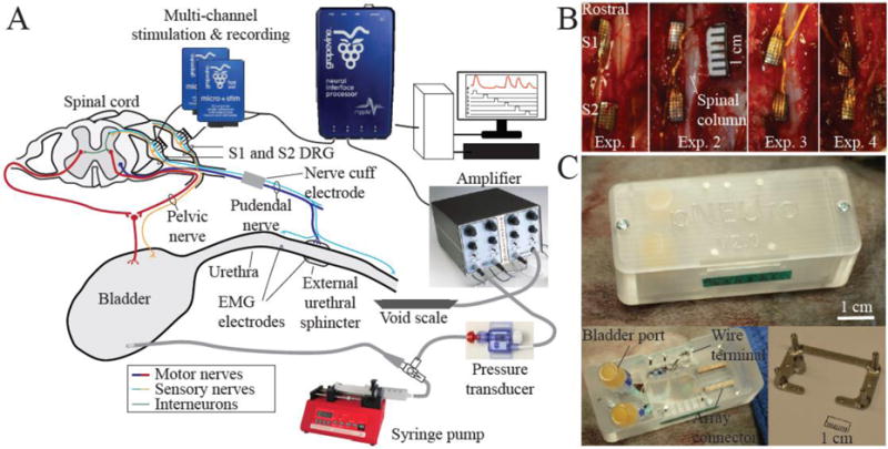
Figure 1.

A. Experimental setup adapted from Bruns et al. 2015 [12]. B. Dorsal view of penetrating microelectrode arrays implanted unilaterally in left S1 and S2 DRG in four felines. C. Custom acrylic backpack housing (top) based on previous designs [42,43]. The backpack with cover removed (bottom left). Bladder catheter ports, array connectors, and cuff and other wires are housed inside the backpack. The stainless steel base plate (bottom right) is mounted on the iliac crest of the animal. The plastic housing is screwed into the four transcutaneous posts of the base plate.
