Fig. 3.

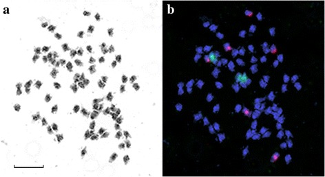
Metaphase plate of L. marginale 2n = 84. a – inverted DAPI staining of chromosomes (grey). b – FISH with labeled 35S (green) and 5S (red) rDNA probes. Scale bar – 5 μM

Metaphase plate of L. marginale 2n = 84. a – inverted DAPI staining of chromosomes (grey). b – FISH with labeled 35S (green) and 5S (red) rDNA probes. Scale bar – 5 μM