
Fig. 1.
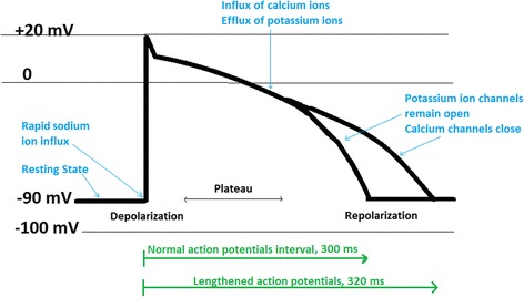
Schematic changes in ventricular action potential at the molecular level within the cardiomyocytes. The resting membrane potential of cardiomyocytes is about −90 mV (mV) while at the full depolarization it can be gradually shifted to +20 mV. In the repolarization stage, membrane potential will return to −90 mV. Some drugs can prolong the duration of normal action potential (lengthened action potential in green) which eventually can lead to drug-induced arrhythmia. Consequently, production of lengthened action potential (long QT syndrome) may initiate TdP arrhythmia (Adapted with permission from [31])