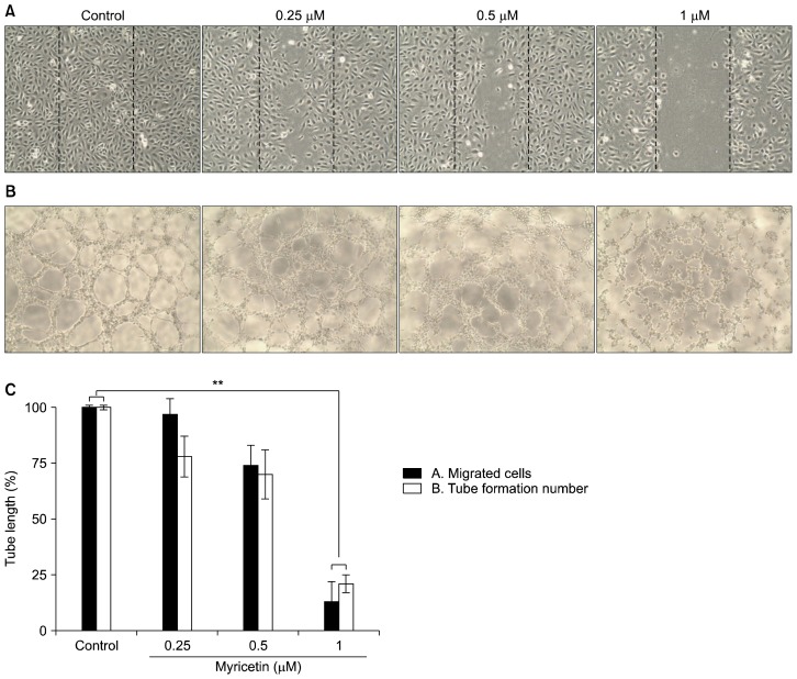
Figure 4.
Effects of myricetin on migration and tube formation in human umbilical vascular endothelial cells (HUVECs). (A) HUVECs were grown to confluence in 6-well plates, scratch-wounded, and treated with the indicated concentrations of myricetin. Cell migration was visualized under an optical microscope (×100). (B) HUVECs were cultured in 96-well plates coated with Matrigel and incubated for 4 to 8 hours in the absence or presence of myricetin (×100). (C) The numbers of migrated cells and tube formations in HUVECs after myricetin treatment were counted; values represent the mean ± SD; **P < 0.01 compared with control.

