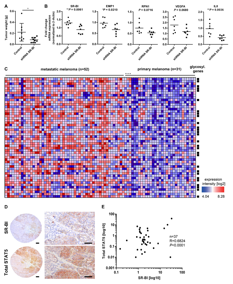
Figure 6. SR-BI promotes melanoma progression and its signature classifies melanoma in metastatic and non metastatic groups.
(A) Tumor weights after intradermal transplantation of 451Lu* cells either stable transduced with control shRNA (n=8) or shRNA targeting SR-BI (n=10) into SCID mice. (B) Tumor mRNA was isolated and RT-PCR of indicated genes was performed (n=7 per group). (C) Enrichment displayed as a heat map of SR-BI target genes in the patient dataset GSE8401 classifies primary and metastatic melanoma patients. Squares symbols indicate genes involved in the glycosylation pathway. (D) Immunohistological stainings of consecutive tissue from microarray slides anti-SR-BI and anti-total STAT5. Scale bar, 50µm. (E) Correlation analysis of SR-BI with STAT5 in metastatic samples derived from patient lymph nodes.

