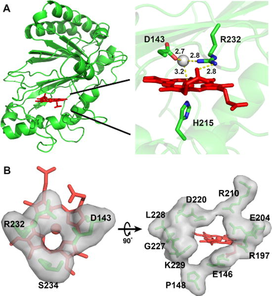
Figure 2.

Crystal structure of wt-ElDyP. (A) Overall structure (left) and active site of the enzyme. Catalytic residues, heme, and water 288 are represented in green sticks, red sticks, and a grey ball, respectively. Distances in angstrom are labeled and shown in yellow dashed lines. (B) Surface representations of the small (left, diameter of ~3.0 Å) and large (right, diameter of ~8.0 Å) heme access channels.
