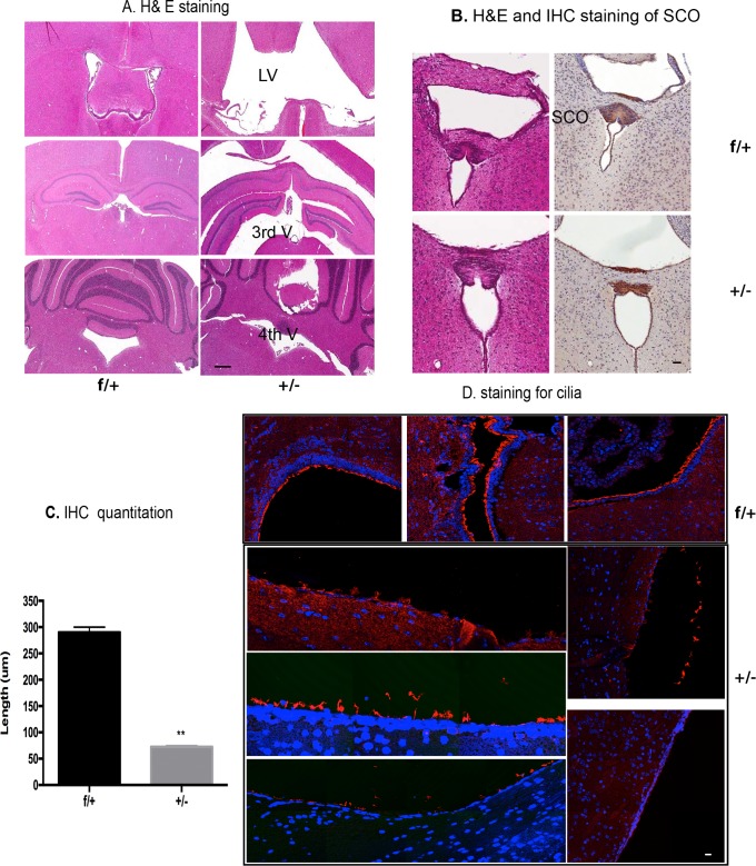
Fig 2. Characterization of Rfx4+/- mice with hydrocephalus.
(A) H&E staining. The left panels are representative slides from a control mouse at 4 weeks of age (Rfxf/+), and the right panels were from a Rfx4+/- mouse, with roughly equivalent sections shown descending from rostral to caudal. The sections demonstrate the enlarged lateral and third ventricles, as indicated by the arrows, but not fourth ventricle enlargement, in the Rfx4+/- mouse (representative of 6 Rfx4+/- mice and 4 controls). The middle right panel shows the emergence of false ventricles, and the white matter appeared to be compressed from the severe hydrocephalus. Scale bar = 20 μm. (B) SCO hypoplasia in Rfx4+/- mice. The left panels of B showed the SCO stained with H&E, with a section from a control mouse (Rfxf/+) on top, and from an Rfx4+/- KO mouse on the bottom. Neighboring sections were stained with an antibody to Reissner’s fibers, and the resulting immunohistochemistry is shown on the right. Scale bar = 10 μm. (C) IHC quantitation. SCO length was measured by counting positively stained Reissner’s fibers slides from 3 controls and 3 Rfx4+/- mice, using adjacent sections cut 5 μm apart. These measurements showed that SCO length in Rfx4+/- KO mice was decreased when compared to controls (p<0.01). (D) Immunofluorescence staining. Acetylated alpha-tubulin labeled cilia (red fluorescence, blue is DAPI) were detached, patchy or missing from the walls of the ventricles in Rfx4+/- mice (lower five panels). Scale bar = 5 μm.

