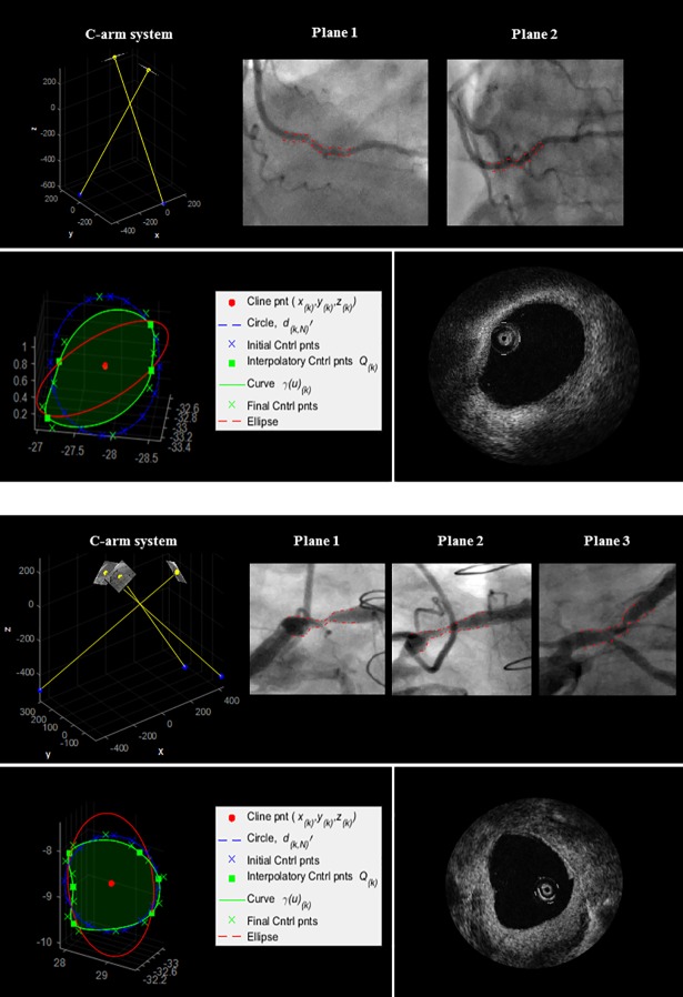
Fig 10. 3D reconstruction of Patient 1 and Patient 2 (Table 2).
Top: 2D angiographic images and their 3D relationship. Bottom: an example of 3D reconstructed luminal cross-section (green), circular fitting (blue), and elliptical fitting (red); corresponding OCT frame.

