
Figure 2. In vitro neurite growth 7 days after in vivo ES of the sciatic nerve.
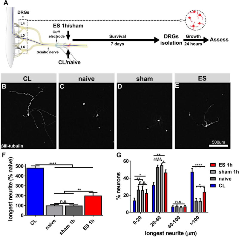
(A) Experimental design. DRGs (L4-L6) were isolated from (B) animals 7 days after CL, (C) naïve animals, (D) 7 days after sham or (E) after electrical stimulation. Cells were grown on PDL-laminin (5μg/ml) coated plates for 24 hours and labeled for βIII-tubulin. (F) Quantification of neurite growth demonstrates a strong effect of conditioning lesions, but 1h ES 7d prior to isolation is also growth-promoting (n=8/group, ANOVA p<0.0001, Tukey´s posthoc test). (G) Neurons classified by their longest neurites reveal that conditioning lesions enhance neurite initiation and extension, whereas electrical stimulation primarily enhances neurite extension (n=8/group, ANOVA p<0.0001, Tukey´s posthoc test). Mean ± SEM. *p<0.05, **p<0.01, ***p<0.001, ****p<0.0001.