
Figure 1. Isolation of cilia from sea urchins, sea anemones and choanoflagellates.
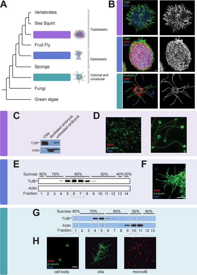
(A) The phylogenetic relationship of the organisms studied in this work to other eukaryotes. (B) Immunofluorescent staining of cilia, marked by β-tubulin or Tubulinac (TUBac) (green), in a sea urchin (Strongylocentrotus purpuratus) gastrula embryo, a sea anemone (Nematostella vectensis) planula larva, and a colony of choanoflagellate cells (Salpingoeca rosetta). Phalloidin staining of the sea anemone larva demonstrates the localization of Actin (red) to the cell bodies. Phalloidin staining of the choanoflagellate colony marks the collar of microvilli that surrounds each flagellum. Nuclei are stained with DAPI (blue). (C) Lysates of isolated S. purpuratus embryonic cilia, deciliated embryos, and intact embryos immunoblotted for TUBac and Actin. Cilia are enriched for TUBac and have undetectable amounts of Actin, whereas deciliated embryos have undetectable amounts of TUBac. (D) Immunostaining of purified S. purpuratus cilia for β-tubulin (green), Actin (red) and nuclei (DAPI, blue) demonstrates that the axonemes of isolated cilia remain intact (β-tubulin) and confirms that no Actin or nuclei are detected in the ciliary fraction. (E) Immunoblot analysis of fractions from the sucrose step gradient purification of isolated N. vectensis cilia reveals that the 60% sucrose fractions contains TUBac, peaking in fractions 6 and 7. (F) Immunostaining of fraction 7 for β-tubulin (green), Actin (red) and nuclei (DAPI, blue) confirms that the sea anemone ciliary fraction is replete with cilia (TUBac) and contains little Actin and no detectable nuclei. (G) Immunoblot analysis of fractions from the sucrose step gradient purification of S. rosetta components reveal that TUBac is enriched in the 70% sucrose step and Actin is enriched in the 50% sucrose step. (H) Immunostaining of fractions for β-tubulin (green), Actin (red) and nuclei (DAPI, blue) confirms that cilia are enriched in the 70% sucrose step, cell bodies identified by DAPI staining are in the 80% sucrose step and microvilli marked by Actin are enriched in the 50% sucrose step. Scale bars, 5 μm for all images. (See Table S1).