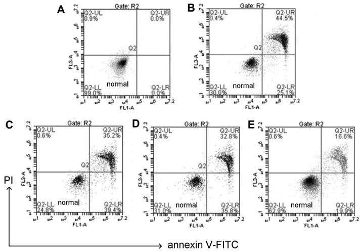
Figure 4.
Apoptosis analysis of human lens epithelial cells. (A) Apoptosis in blank cells without any treatment, or pretreated with different concentration of chlorogenic acid: (B) 0 µM, (C) 10 µM, (D) 30 µM, (E) 50 µM, followed by 100 µM H2O2 for 24 h. Apoptosis was analyzed using flow cytometry with Annexin V/PI staining. Control cells were treated with H2O2 (100 µM) alone. P<0.01 control group vs. all other groups. PI, propidium iodide; FITC, fluorescein isothiocyanate.

