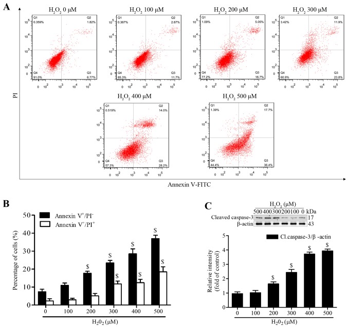
Figure 2.
H2O2 induces apoptosis of MSCs. (A and B) Cells were treated with a range of H2O2 concentrations for 4 h, and apoptosis was analyzed by Annexin V-FITC/PI staining assay. (C) H2O2-induced cleaved caspase-3 expression was measured by western blotting, β-actin was used for normalization. Data are presented as the mean ± standard deviation of three independent measurements. $P<0.05 compared with the 0 µM H2O2 group. Cl.caspase-3, cleaved caspase-3; FITC, fluorescein isothiocyanate; H2O2, hydrogen peroxide; MSCs, mesenchymal stem cells; PI, propidium iodide.

