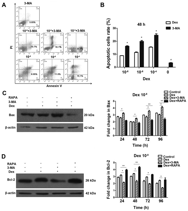
Figure 4.
Regulation of autophagy interferes in the negative effects of Dex on MC3T3-E1 cells. (A) MC3T3-E1 cells were treated with Dex, in the presence or absence of chemical inhibitors of autophagy 3-MA. Annexin V-fluorescein isothiocyanate/PI staining was analyzed by flow cytometry. Representative dot plots of MC3T3-E1 cells subjected to Dex or 3-MA alone are presented. (B) Data expressed as percentage of Annexin V-positive cells (n=3). *P<0.05, vs. corresponding control and #P<0.05 other groups vs. 3-MA alone. Cells were treated with 10−8 mol/l Dex and 3-MA (400 μmol/l) or RAPA (500 nmol/l), and the expression of (C) Bax and (D) Bcl-2 were detected by western blotting. The results were representative of three independent experiments. β-actin was used as a loading control. *P<0.05; **P<0.05. 3-MA, 3-methyladenine; PI, propidium iodide; Dex, dexamethasone; RAPA, rapamycin; Bax, BAX apoptosis regulator; Bcl-2, Bcl-2 apoptosis regulator.

