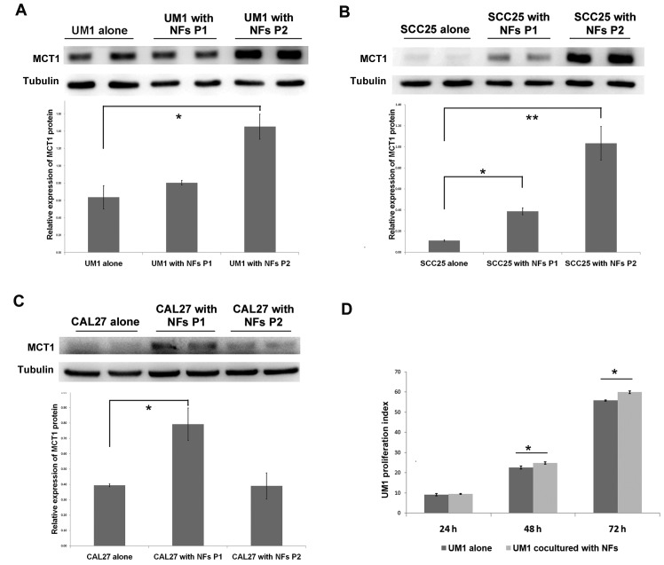
Figure 6.
Fibroblasts promote MCT1 expression and proliferation of OSCC cells. OSCC cell lines (A) UM1, (B) SCC25 and (C) CAL27 and normal NFs were co-cultured for two passages. MCT1 expression of OSCC cell lines was detected by western blot assay. Densitometry was used to determine MCT1/tubulin ratios. *P<0.05. (D) Carboxy-fluorescein succinimidyl ester pre-stained UM1 and NFs were co-cultured for 72 h, and these UM1 cells were collected and fixed every 24 h until flow cytometry analysis. The histogram presented the fluorescence signals at 488 nm exciting light and proliferation index (represent the mean ± standard deviation of three independent experiments) was calculated according to the signal profile. *P<0.05 and **P<0.01. OSCC, oral squamous cell carcinoma; NFs, normal fibroblasts; P, passage; MCT1, mono-carboxylate transporter 1.

