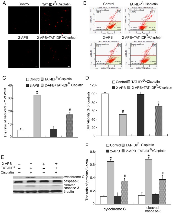
Figure 5.
2-APB reduces the growth inhibition and apoptosis induced by TAT-IDPS and cisplatin. SKOV3/DDP cells were treated with 15 μg/ml cisplatin and 25 μM TAT-IDPS in the presence or absence of 50 μM 2-APB for 24 h. (A) Cells were incubated with the fluorescent calcium indicator Rhod-2/AM and mitochondrial Ca2+ levels were observed by confocal microscopy (scale bar, 40 μm). (B) ΔΨm was assessed by staining with MitoPotential dye and 7-aminoactinomycin D, and was analyzed using the Muse® Cell Analyzer. (C) Quantification of ΔΨm. Data are presented as the mean ± standard deviation, n=3. (D) Cell viability was determined using an MTT assay. Data are presented as the mean ± standard deviation. (E) Western blot analysis of the expression of cytochrome c and cleaved caspase-3 in SKOV3/DDP cells. (F) Semi-quantification of cytochrome c and cleaved caspase-3 expression. Data are presented as the mean ± standard deviation, n=3. *P<0.05 vs. the control group; #P<0.05 vs. the cisplatin + TAT-IDPS group. ΔΨm, mitochondrial membrane potential; 2-APB, 2-aminoethyl diphenylborinate; TAT-IDPS, TAT-fused IP3R-derived peptide.

