. 2018 Jan 3;7:e32273. doi: 10.7554/eLife.32273

Figure 2. The tandemly duplicated bab genes perform a derived role in repressing a CRE controlling male-specific expression of the gene yellow.

(A) An ancestral bab gene was duplicated into the paralogous bab1 and bab2 genes in a Dipteran lineage that includes Drosophila fruit flies. The time scale indicates approximate divergence times in millions of years ago. (B) Male-specific expression of yellow in the abdominal epidermis is under the control of the yBE0.6 CRE that possesses two binding sites for Abd-B that are shown as yellow rectangles. Blue bars delimit the SM4 and SM10 regions required to suppress CRE activity in females. (C and D) The yBE0.6 EGFP reporter transgene is elevated in the male A5 and A6 abdomen segments (C) but is only barely detected females (D). (E–G) Ectopic reporter expression occurs in the female abdomen when either the SM4, SM10, or both regions are mutated. (H) The pnr-GAL4 driver activates dorsal midline expression of the UAS-EGFP gene, demarcating its domain of misexpression. (I) Dorsal midline expression of the yBE0.6 CRE is lost when bab1 is ectopically expressed by pnr-GAL4. (J) When the SM4 and SM10 regions are mutated, the yBE0.6 CRE can activate reporter expression in midline regions in spite of ectopically expressed bab1.

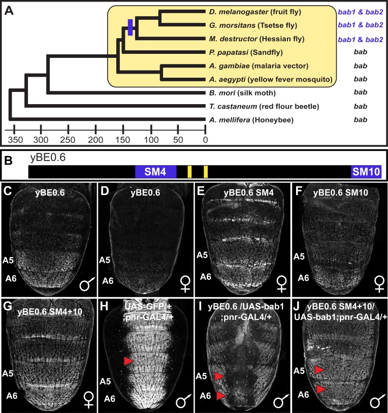
Figure 2—source data 1. Amino acid alignment for Bab homologs.
The amino acid sequences for Drosophila (D.) melanogaster Bab1 and Bab2, D. ananassae Bab1 and Bab2, D. willistoni Bab1, D. mojavensis Bab2, Glossina morsitans Bab1 and Bab2, and Anopheles gambiae Bab were aligned using the Clusta Omega multiple sequence alignment program. The BTB and Conserved Domains for D. melanogaster Bab1 and Bab2 are respectively indicated by the maroon and blue background colors. Within the Conserved Domain the psq domain is indicated by the amino acids with yellow font color and the AT-hooks by amino acids with red font color.
DOI: 10.7554/eLife.32273.005
Figure 2—source data 2. Sequence alignment of the yBE0.6 with scanning mutant versions.
Purple background with white letters indicate the AscI (GGCGCGCC) and SbfI (CCTGCAGG) restriction enzymes sites that were appended to primers for cloning CRE versions into matching sites in the S3aG reporter transgene vector. Maroon background and white letters indicate sequences that comprise scanning mutations. The lower case nucleotide letters indicate the non-complementary transversions. The yellow background with black letters indicates the Abd-B sites identified in Jeong et al. (2006) which were not mutated in this study. The blue background with white letters indicate the Bab-bound sequences identified in this study. The gray background with bolded black letters indicate the regions to which the BE2.5 Fwd and BE3.5 Rvs primers (reverse complement of that highlighted) were designed to initially amplify the wild-type CRE sequence.
DOI: 10.7554/eLife.32273.006

Figure 2.

Figure 2—figure supplement 1. Scanning mutagenesis across the entire yBE0.6 CRE identifies sequences that normally function to repress CRE activity in the female abdomen.

Figure 2—figure supplement 1.

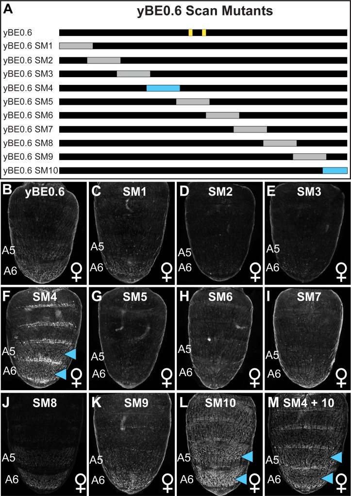
(A) Name and location of yBE0.6 scanning mutations. Scanning mutations for which CRE activity in the female abdomen was not noticeably altered are indicated as gray rectangle and those for which ectopic activity occurred are shown as light blue rectangles. The two vertical yellow lines on the illustration of the wild type CRE indicate the position of previously identified Abd-B binding sites that were not mutated in this study. (B–M) The EGFP reporter gene expression pattern in the female abdomen at ~85 hr after puparium formation driven by the non-muntant (yBE0.6) and scan mutant CRE sequences. (M) Scanning mutations 4 and 10 were combined together. Light blue arrowheads indicate abdomen segments with conspicuous ectopic EGFP expression.