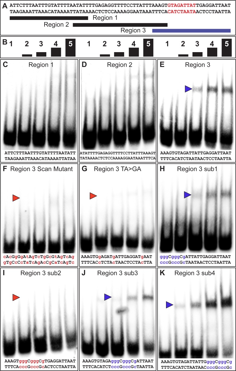
Figure 3. The yBE0.6 possesses a binding site for Bab in the SM4 region.
(A) The wild type DNA sequence of the SM4 region is shown, which was subdivided into three smaller regions annotated below that were used as double stranded probes in gel shift assays with the GST-Bab1 DNA-binding Domain (Bab1-DBD). Red text delimits the inferred Bab-binding site. (B) Each probe was tested in gel shift assay reactions for binding with five different amounts of Bab1-DBD. These were from left to right: 0, 500, 1000, 2000, and 4000 ng. (C–E) Gel shift assays using wild type probe sequences. (F–K) Gel shift assays using mutant probe sequences. Lower case blue letters indicate probe mutations that did not noticeably alter protein binding. Probe base pairs in lower case red letters are changes that altered protein binding. Blue and red arrowheads indicate the location of shifted probe, with red arrowheads indicating cases where the quantity of shifted probe was noticeably reduced.

