. 2018 Jan 3;7:e32273. doi: 10.7554/eLife.32273

Figure 6. Bab1 and Bab2 are sufficient to suppress tergite pigmentation as DNA-binding transcription factors.

(A–L) Ectopic expression assays for various bab protein coding sequences. (A and G) D. melanogaster bab1, (B and H) D. melanogaster bab2, (C and I) a DNA-binding compromised version of D. melanogaster bab1 (bab1 DBM), (D and J) D. willistoni bab1, (E and K) D. mojavensis bab2, and (F and L) A. gambiae bab. (A–F) Leaky expression of transgenes from the attP40 transgene insertion site. (G–L) Ectopic expression of protein coding sequences in the male abdomen under the control of the y-GAL4 transgene. Red arrowheads indicate tergite regions with conspicuously reduced tergite pigmentation. The A5 and A6 tergite regions were quantified for their darkness percentage for replicate specimens (n = 4). These percentages and their standard error of the mean (±SEM) are provided below a representative image.

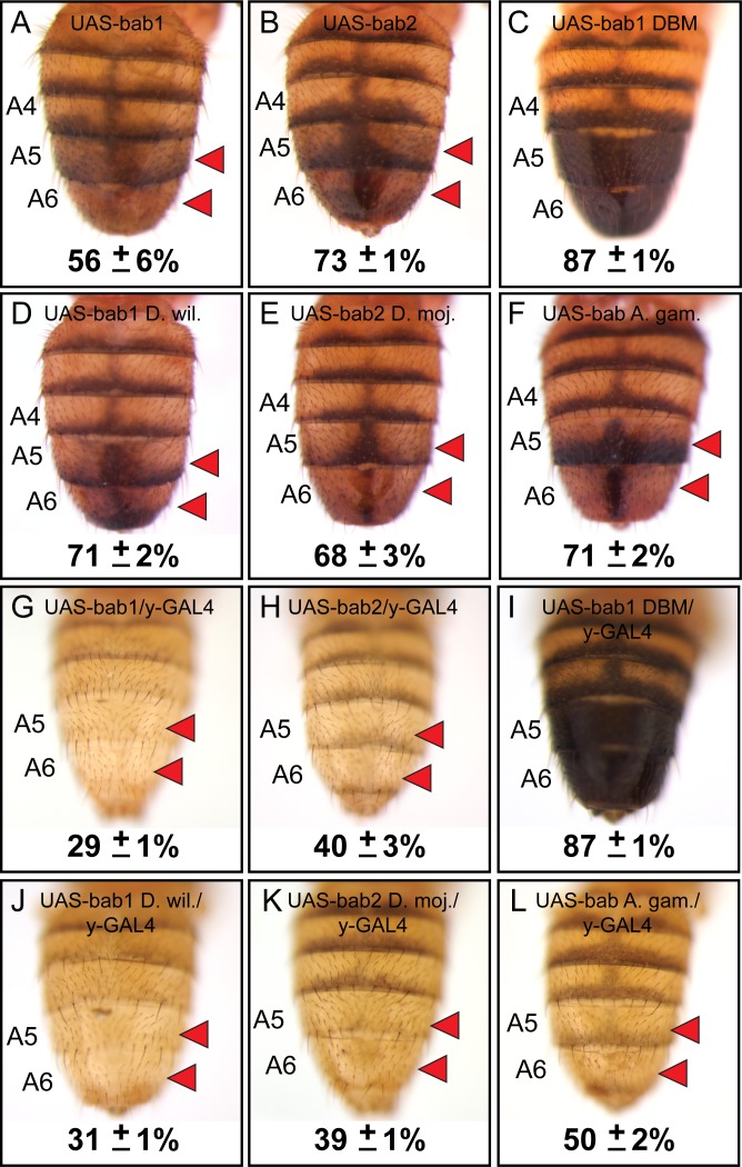
Figure 6—source data 1. The DNA and translated protein sequences for the bab open-reading frames.
DOI: 10.7554/eLife.32273.018
Figure 6—source data 2. Analysis of pigmentation phenotypes from bab open-reading frame ectopic expression.
DOI: 10.7554/eLife.32273.019

Figure 6.

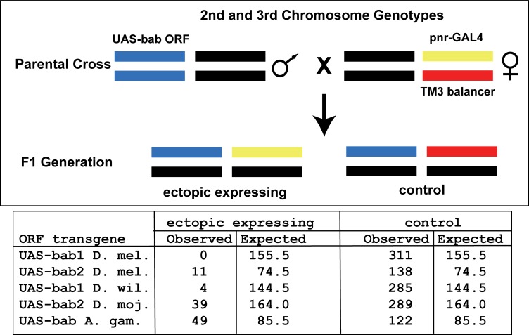
Figure 6—figure supplement 1. Lethality from ectopic expression of orthologous bab open reading frame transgenes in the pnr pattern.

Figure 6—figure supplement 1.

UAS-bab open-reading frame transgenes are located in the attP40 site on the D. melanogaster second chromosome. Male flies were crossed to females heterozygous for the third chromosome where the GAL4 gene is inserted into the pnr locus and the TM3 balancer. Fewer offspring were obtained that possessed an ectopic bab expressing genotype than expected by chance, indicating lethality due to bab expression in the spatial and temporal pattern of the pnr gene.
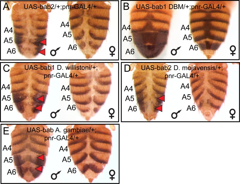
Figure 6—figure supplement 2. Bab proteins are sufficient to suppress tergite pigmentation when ectopically expressed in the dorsal midline of D. melanogaster.

Figure 6—figure supplement 2.

(A–E) Ectopic expression of bab protein coding sequences in the dorsal midline of male and female abdomens under control of the pnr-GAL4 driver. These coding sequences were (A) D. melanogaster bab2, (B) a DNA-binding compromised version of D. melanogaster bab1 (bab1-DBM), (C) D. willistoni bab1, (D) D. mojavensis bab2, and (E) A. gambiae bab. Red arrowheads indicate tergite regions with conspicuously reduced tergite pigmentation. (A, C, D, and E) Ectopic expression resulted in reduced tergite pigmentation and a non-specific split tergite phenotype. (B) The DNA-binding mutant Bab1 could neither suppress tergite pigmentation nor cause the non-specific split tergite phenotype.
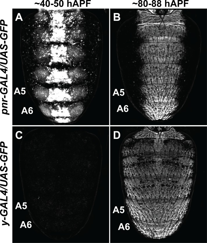
Figure 6—figure supplement 3. The temporal and spatial domains of activity for GAL4 drivers in D. melanogaster pupa.

Figure 6—figure supplement 3.

(A) Dorsal midline expression of the UAS-GFP transgene under the control of the pnr-GAL4 driver at ~40–50 hr after puparium formation (hAPF). (B) Dorsal midline expression of the UAS-GFP transgene under the control of the pnr-GAL4 driver at ~80–88 hAPF. (C) The UAS-GFP transgene is not expressed at ~40–50 hAPF when under the regulatory control of the y-GAL4 driver. (D) The pan-abdomen expression of the UAS-GFP transgene under the control of the y-GAL4 driver at ~80–88 hAPF. The highest level expression occurs in the A5 and A6 segments due to the activity of the body element CRE which is included in the y-GAL4 transgene. All specimens shown are males.
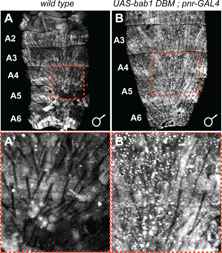
Figure 6—figure supplement 4. Ectopic expression of the Bab1-DNA-binding mutant protein.

Figure 6—figure supplement 4.

(A) Little-to-no endogenous Bab1 protein can be detected in the dorsal abdominal epidermis of D. melanogaster male pupa. (B) In contrast, nuclear-localized expression of the Bab1 DNA-binding mutant protein can be observed when the UAS-transgene was ectopically expressed under the control of the pnr-GAL4 driver. (A’ and B’) Zoomed in view of the expression within the regions outlined by dashed red boxes in panels A and B. Samples shown are at a pupal developmental stage of ~88 hr after puparium formation.