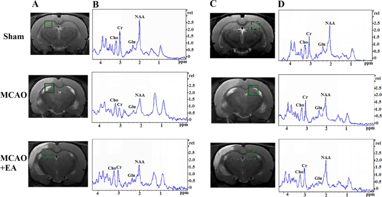
Figure 4.
Changes in brain metabolites in HPC by MRS in rats with cerebral I/R injury. (A and C) Localization of the left and right HPC VOI on the T2-weighted scan, which is an MRS shimming region. (B and D) 1H-MRS exhibited the NAA peak at 2.02 ppm, the Glu peak at 2.2 ppm, the Cho peak at 3.20 ppm, and the Cr peak at 3.05 ppm. HPC, hippocampus; MRS, magnetic resonance spectroscopy; I/R, ischemia and reperfusion; VOI, volume of interest; NAA, N-acetylaspartate; Glu, glutamate; Cho, choline; Cr, creatine.

