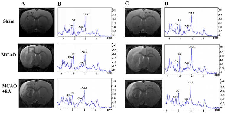
Figure 5.
Changes in brain metabolites in TPC by MRS detection in rats rats with cerebral I/R injury. (A and C) Localization of the left and right TPC VOI on the T2-weighted scans, which is an MRS shimming region. (B and D) 1H-MRS exhibited the NAA peak at 2.02 ppm, the Glu peak at 2.2 ppm, the Cho peak at 3.20 ppm, and the Cr peak at 3.05 ppm. MRS, magnetic resonance spectroscopy; I/R, ischemia and reperfusion; VOI, volume of interest; NAA, N-acetylaspartate; Glu, glutamate; Cho, choline; Cr, creatine.

