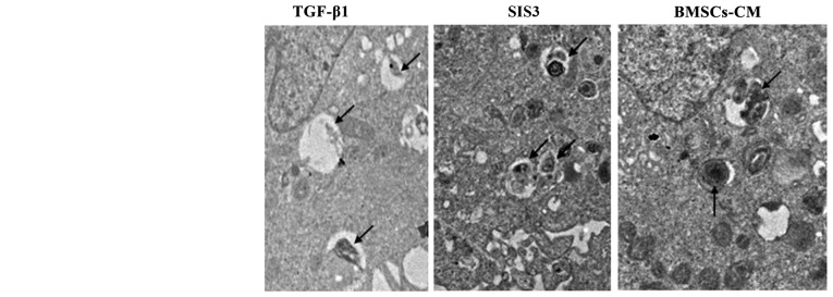
Figure 3.
Ultrastructural evidence of EMT. Normal A549 cells can be identified by the presence of LBs in the cytoplasm. In the TGF-β1 group, LBs were degeneration, swollen and with vacuoles. Arrows indicate LBs; magnification, ×7,000. The cell groups were as follows: control, cells cultured in serum-free DMEM; TGF-β1, cells cultured in serum-free DMEM and exposed to 5 ng/ml TGF-β1; SIS3, cells were treated with 3 μM SIS3 (specifc inhibitor of Smad3), which was added 4 h prior to 5 ng/ml TGF-β1 exposure; BMSCs-CM, BMSCs-CM was added prior to 5 ng/ml TGF-β1 exposure. Nu, cell nucleus; LB, lamellar body; EMT, epithelial-mesenchymal transition; TGF-β1, transforming growth factor-β1.

