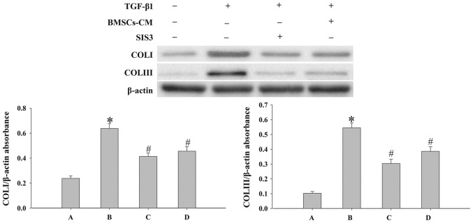
Figure 8.
Evaluation of the expression levels of COLI and COLIII in cells. Western blot analysis was used to examine the levels of COLI and COLIII in the cells. Densitometric analysis of protein expression with β-actin as the control. The results demonstrated that treatment with BMSCs-CM or SIS3 significantly decreased the expression of COLI and COLIII following exposure to TGF-β1 (*P<0.05 vs. control group; #P<0.05 vs. TGF-β1 group). The bars are labeled as follows: A, control group; B, TGF-β1 group; C, SIS3 group; and D, BMSCs-CM group. The cell groups were as follows: control, cells cultured in serum-free DMEM; TGF-β1, cells cultured in serum-free DMEM and exposed to 5 ng/ml TGF-β1; SIS3, cells were treated with 3 μM SIS3 (specific inhibitor of Smad3) which was added 4 h prior to 5 ng/ml TGF-β1 exposure. BMSCs, bone marrow-derived mesenchymal stem cells; CM, conditioned medium; TGF-β1, transforming growth factor-beta 1; COLI, collagen I.

