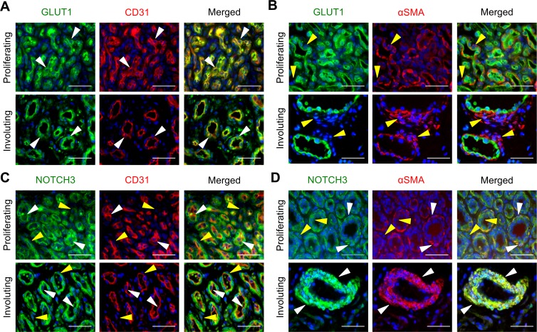
Figure 1. NOTCH3 is expressed in perivascular and lumenal cells in IHs.
Serial sections of proliferating and involuting infantile hemangioma (IH) specimens were stained. (A) GLUT1 and CD31 costaining. White arrowheads mark GLUT1+CD31+ cells. Proliferating IH n = 2, involuting IH n = 3. (B) GLUT1 and αSMA costaining. Yellow arrowheads mark αSMA+GLUT1– perivascular cells. Proliferating IH n = 2, involuting IH n = 7. (C) NOTCH3 and CD31 costaining. White arrowheads mark NOTCH3+CD31+ cells. Yellow arrowheads mark NOTCH3+CD31– cells. Proliferating IH n = 4, involuting IH n = 5. (D) NOTCH3 and αSMA costaining. White arrowheads mark NOTCH3+αSMA+ perivascular cells. Yellow arrowheads mark NOTCH3+αSMA– lumenal cells. Proliferating IH n = 5, involuting IH n = 8. Scale bars: 50 μm. The total number of IH specimens assessed for each antigen is presented in Supplemental Table 3. αSMA, α smooth muscle actin; GLUT1, glucose transporter 1.

