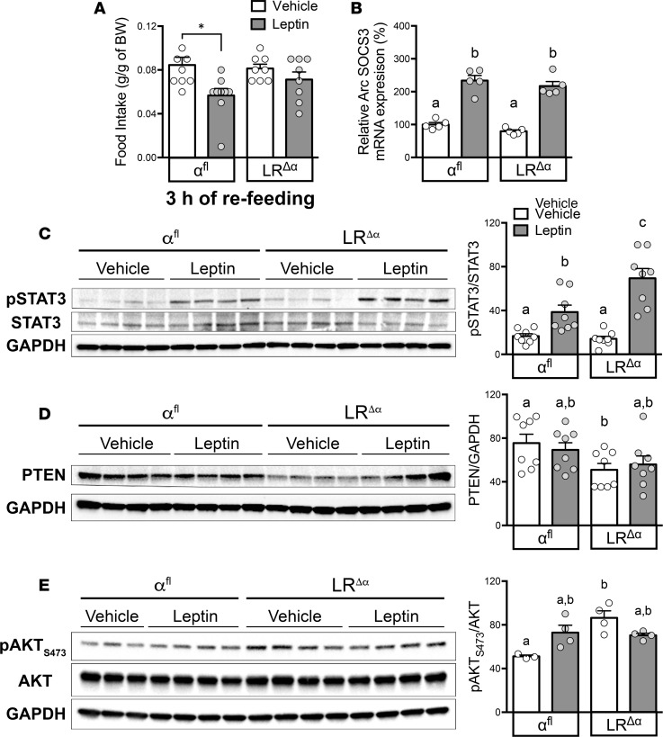
Figure 6. LRΔα mice show enhanced leptin sensitivity and reduced hypothalamic PTEN expression.
(A) Blunted acute anorexigenic effect of leptin (3 hours of refeeding) in LRΔα (n = 8/treatment) compared with αfl (n = 9/treatment) males [F(1,30) = 8.89, P = 0.006]. (B) Leptin induced similar SOCS3 mRNA expression in the arcuate nucleus (Arc) of fasted LRΔα and αfl females [n = 5/group; F(1,16) = 150.7, P < 0.0001]. (C) Representative Western blotting and relative protein quantification from hypothalamic extracts show enhanced leptin-induced pSTAT3 in fasted LRΔα compared with αfl (n = 8/treatment, females [F(1,28) = 6.3, P = 0.018 for genotype analysis; F(1,28) = 46.35, P < 0.0001 for treatment analysis]. (D) Reduced PTEN expression from hypothalamic extracts of fasted mice injected with vehicle or leptin [F(1,28) = 7.02, P = 0.013; n = 8/treatment, females]. (E) Representative Western blotting and relative protein quantification of pAKT at Ser473 residue (pAKTS473) from hypothalamus of fasted mice injected with vehicle or leptin (n = 3–4/treatment, females) showed increased pAKTS473 expression in LRΔα females [F(1,11) = 10.1, P = 0.009]. Each point represents 1 individual mouse. Data are presented as mean ± SEM. *P < 0.05 versus control mice; groups with different superscript letters are statistically different by 2-way ANOVA with Tukey’s post-hoc analysis.

