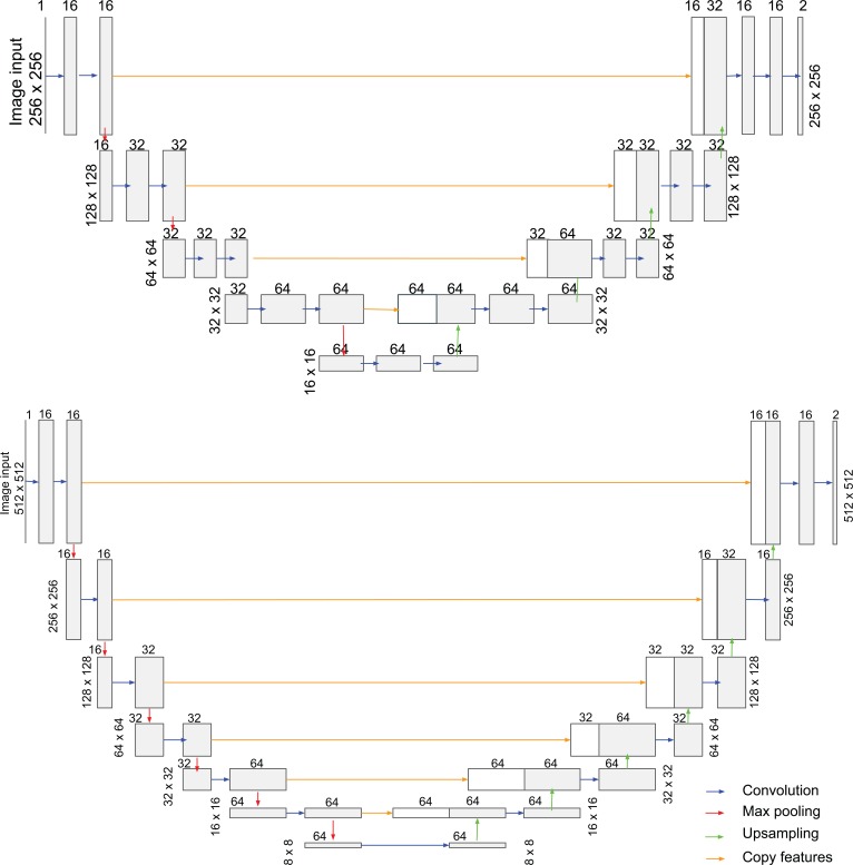
Figure 2. Deep learning model architectures used for vaso-obliteration and neovascular segmentation.
The U-net architecture for vaso-obliteration (VO) segmentation (top) and neovascularization (NV) segmentation (bottom). For each convolutional layer, the filter size is 3 and the stride is 1. The number of filters is labeled on top of its corresponding layer. ReLU was used as the activation function. The receptive fields were 140 × 140 and 318 × 318 for VO and NV segmentation, respectively.

