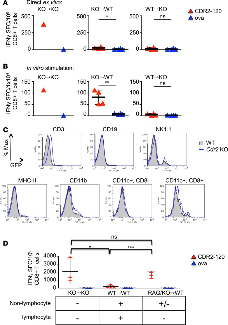
Figure 3. T cell expression of CDR2 is sufficient to confer tolerance.
IFN-γ ELISPOTs of CD8+ T cells from bone marrow (BM) chimera mice 14 days after immunization with adenovirus expressing CDR2 (AdV-CDR2) and cultured with RMA-S cells pulsed with either OVA or CDR2-120 peptide immediately ex vivo (A) and after 7 days of splenocyte in vitro stimulation (B). Each triangle represents the mean of triplicate wells and the bar with error bars represents the mean and standard deviations of mice in that group. These data are representative of 2 experiments. KO→KO indicates Cdr2-KO BM donor cells transplanted into Cdr2-KO host mice (n = 1), KO→WT (n = 4) indicates Cdr2-KO BM donor cells transplanted into WT host mice, and WT→KO (n = 4) indicates WT BM donor cells transplanted into Cdr2-KO host mice. (C) Flow cytometry of hematopoietic cells from WT or Cdr2-KO mice. The EGFP gene replaces the Cdr2 gene in Cdr2-KO mice. Gray filled = WT, blue line = Cdr2-KO. One experiment is shown and is representative of 5 experiments. (D) BM chimeras were established as in A and B with the addition of RAG/Cdr2-KO→WT (n = 3) indicating a mix of 50:50 Rag1-KO and Cdr2-KO BM cells transplanted into WT mice. Fourteen days after immunization, splenocytes were stimulated in vitro for 7 days with CDR2-120 peptide and CD8+ T cells were tested for response to RMA-S cells pulsed with either OVA or CDR2-120 peptide by IFN-γ ELISPOT in triplicate wells. Results presented are 1 of 2 experiments. Each triangle represents the mean of triplicate wells and the bar with error bars represents the mean and standard deviations of mice in that group. These data are representative of 2 experiments. KO→KO (n = 3), WT→WT (n = 5), RAG/KO→WT (n = 3). *P < 0.05, **P < 0.01, ***P < 0.001; ns, statistically not significant as calculated using unpaired Student’s t test. SFC, spot-forming cells; OVA, ovalbumin peptide.

