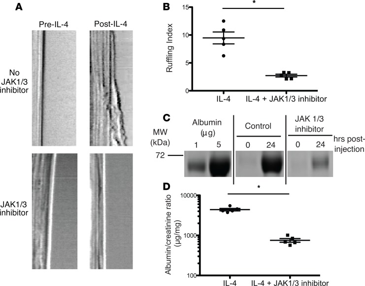
Figure 4. Inhibition of IL-4 signaling with JAK1/3 inhibitor abrogated ruffling and proteinuria in IL-4–treated mice.
(A) Representative kymographs obtained by membrane ruffling and (B) quantification, as performed in Figure 1, from cultured podocytes treated with the JAK1/3 inhibitor tofacitinib (100 nM, bottom row), which significantly attenuated IL-4–induced membrane ruffling compared with negative control (DMSO). Each symbol represents the average relative cell membrane displacement over time at 5 regions of a single cell. Mean ± SD of 2 experiments, with 2–3 cells/experiment (total of 5 cells analyzed/group). (C) Coomassie blue–stained SDS-PAGE and (D) spot albumin/creatinine ratios of urine from IL-4–treated mice treated with JAK1/3 inhibition, which significantly reduced IL-4–induced proteinuria. Urine was collected 24 hours after plasmid administration. Symbols represent individual mice, and bars represent the mean. Mean ± SEM of 2 experiments, with total of 5 mice/group. *P < 0.008 by 2-tailed Mann-Whitney.

