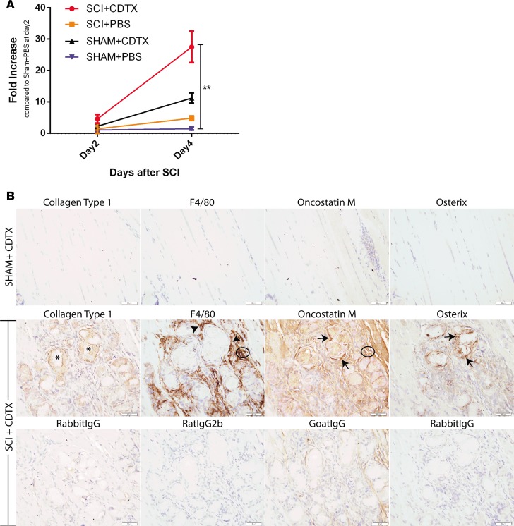
Figure 8. Oncostatin M is expressed at sites of NHO following SCI in mice.
(A) Oncostatin M (Osm) mRNA expression is significantly increased in injured muscle after spinal cord injury (SCI). Total RNA was extracted from right hamstring muscle from mice at days 2 and 4 after SCI or sham surgery and intramuscular cardiotoxin (CDTX) or PBS injection (n = 3/4 mice/group, 1 experiment). Results show qRT-PCR quantification of Osm mRNA (relative to β-actin) with a significant increase in Osm mRNA in muscle 4 days after SCI+CDTX compared with SHAM+PBS mice (**P < 0.01 Kruskal-Wallis test). (B) Representative IHC images of hind limbs from mice that underwent sham or SCI surgery with an intramuscular injection of CDTX. At 21 days after surgery in SHAM+CDTX mice, no neurogenic heterotopic ossification (NHO) is noted in the hamstrings, with few macrophages and absence of osterix or OSM expression. In SCI+CDTX mice, collagen type 1+ (Coll1+) bone foci were noted within the muscle (asterisks); surrounding this NHO are numerous F4/80+ macrophages (arrowheads). IHC for OSM confirmed expression of OSM around areas of NHO, with OSM expression noted in areas of macrophage accumulation (circled area) and osterix+ osteoblasts (arrows). Specificity of staining was confirmed with matched isotype controls (first RabbitIgG: Coll1, RatIgG2b:F4/80, GoatIgG:OSM, and second RabbitIgG:Osterix). Data in A are represented as mean ± SD. Original magnification: ×40. Scale bar: 50 μm.

