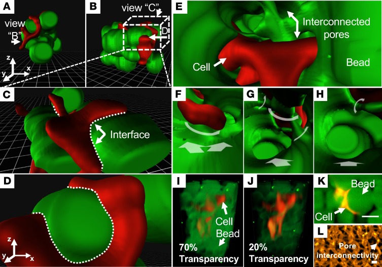
Figure 1. HDFs embedded in a hydrogel matrix.
(A–D) The physical interface between the HDFs (red) and beads (green) provides a user-initiative perception. (E–H) Applying VR-LSFM accentuates the depth perception and contextual relation. Variable stepping directions are labeled with white arrows. (I and J) Conventional 3D volumetric rendering results are constrained to concurrently depict both beads and HDFs due to the reduced levels of transparency of the beads. (K) The 2D raw data reveal attenuation in spatial resolution of the HDF cells interacting with the beads. (L) The polymer network of the 3D microgel matrix is demonstrated in the conventional mode. HDF, human dermal fibroblast; VR, virtual reality; LSFM, light-sheet fluorescence microscopy. Scale bar: 50 μm. All the images are shown in pseudocolor.

