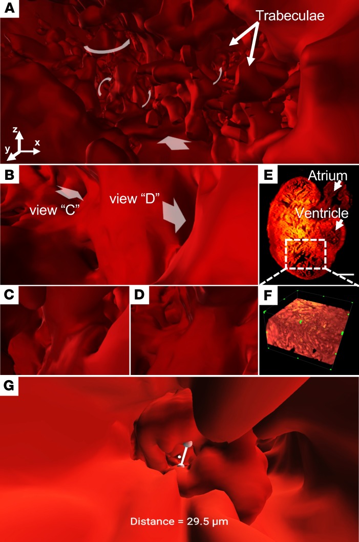
Figure 2. Endocardial trabecular network in a transgenic Tg(cmlc2-gfp) zebrafish ventricle at 60 dpf.
(A) VR accentuates the invaginating muscular ridges in the apical region. (B–D) VR-LSFM enables navigation through various projections into the branching network. Different views are indicated by white arrows (B). (E and F) The conventional (E) 2D raw data and (F) 3D rendering results are limited in revealing the highly trabeculated 2-chambered heart, consisting of an atrium and a ventricle, as the perspective view is predefined. Scale bar: 100 μm. (G) Quantitative measurements of the distance between the ventriculobulbar valve leaflets. dpf, days after fertilization; VR, virtual reality; LSFM, light-sheet fluorescence microscopy. All of these images are shown in pseudocolor.

