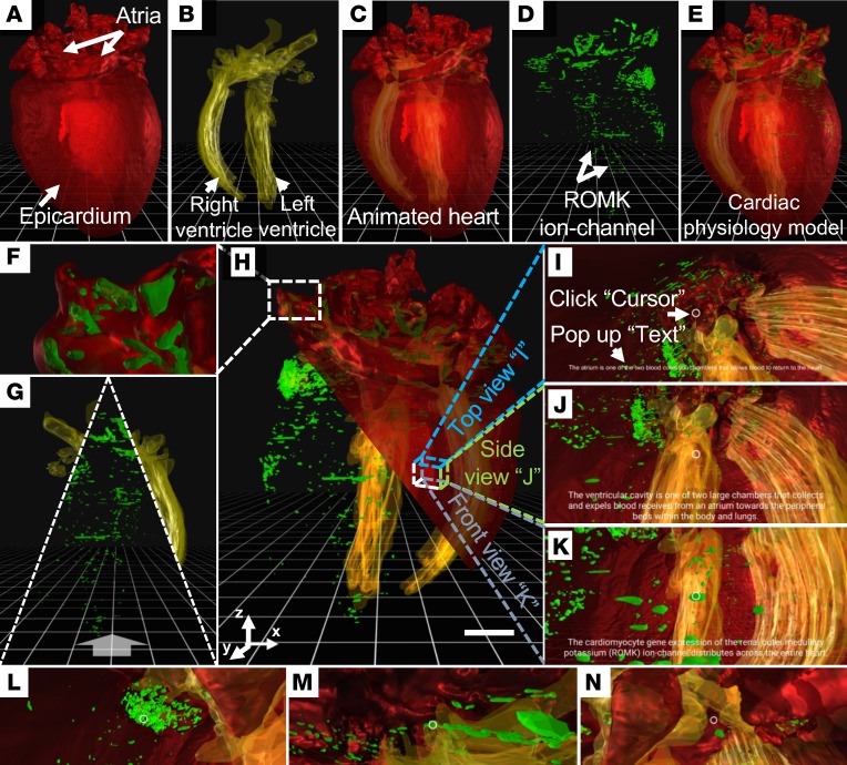
Figure 3. 3D perception of the GFP-labeled ROMK channels in a representative adult mouse heart following gene therapy.
The 3D physical presences of (A) epicardium (red) and (B) ventricular cavity (yellow) are superimposed on the (C) animated heart. (D) 3D distribution of ROMK channels (green) is superimposed with C to provide a (E) physiological model. (F) The distribution of ROMK channels in the atrium. (G) The exploration of the animated ventricular cavity or ROMK channels. (H) This illustration integrates the epicardium (red), ventricular cavity (yellow), and ROMK channels (green) for an interactive and immersive experience. Scale bar: 1 mm. (I–K) VR-LSFM allows for reading instructive texts by clicking the 3D anatomic features of (I) the atrium, (J) the ventricular cavity, and (K) ROMK channels. (L–N) VR-LSFM enables zooming into the 3D anatomy to interrogate ROMK channels in relation to the ventricular cavity and myocardium. ROMK, renal outer medullary potassium; VR, virtual reality; LSFM, light-sheet fluorescence microscopy. All of these images are shown in pseudocolor.

