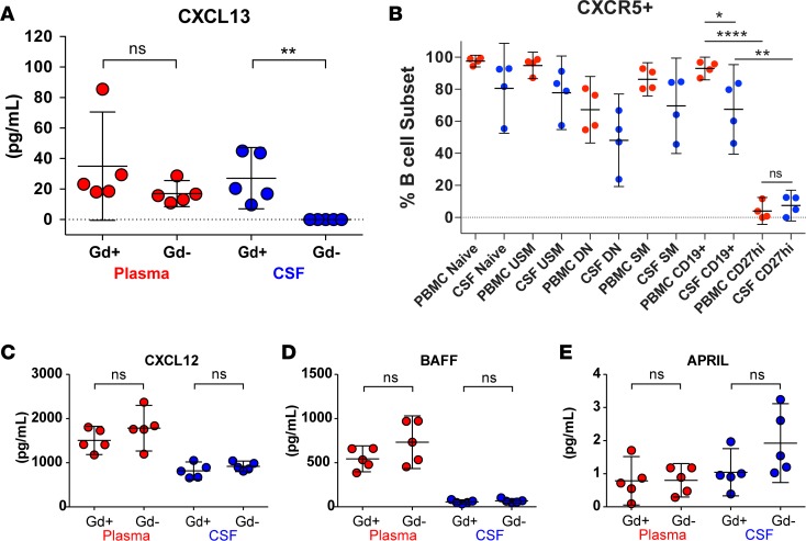
Figure 7. Locally produced CXCL13 may drive B cell recruitment to the CNS.
CXCL13 was undetectable in CSF from Gd– patients, while CXCL13 levels in plasma and Gd+ CSF were similar (A). CXCR5 is expressed on the majority of CSF and PBMC B cells (B). In combination, CXCR5 is present on more PBMCs than CSF CD19+ B cells and only expressed on a minority of PBMCs and CSF CD27hi plasmablasts/plasma cells. There was no significant difference between CXCL12 (C), BAFF (D), and APRIL (E) in CSF from Gd+ or Gd– patients. Overall CXCL12 and BAFF levels were higher in plasma than in CSF. Cytokine/chemokine concentrations were determined in CSF and plasma samples from Gd+ and Gd– patients by ELISA (n = 5 per group; see Table 1). Data are shown as scatter plots with mean ± 95% CI. Comparisons were made using unpaired or paired t tests; *P < 0.05, **P < 0.01, ****P < 0.0001. N, naive B cells; USM, unswitched-memory B cells; DN, double-negative B cells; SM, switched-memory B cells; PC, plasma cells/plasmablasts.

